


What Happens at the 20-Week Ultrasound?

“This one’s got long legs,” the sonographer whispered as she poked my belly with a jelly-covered probe. It wasn’t exactly comfortable laying on that cold exam table, but I felt overjoyed in that moment. Minutes earlier, the technician had told me that I was having a girl.
Reaching the halfway point of pregnancy is a milestone. You’re probably a little less tired, you’re finally showing and you may be feeling baby kick and move around more. It’s also time for your 20-week ultrasound, which gives your ob-gyn or midwife a peek at how baby’s size and overall anatomy are tracking.
It’s normal to feel anxious about the 20-week ultrasound, when the technician will perform an anatomy scan. It’s often an incredibly happy event—but it can sometimes reveal signs that something’s not quite right with your pregnancy or baby. Don’t panic; knowledge is power, and having more intel on what’s happening with you and baby can help your doctor develop an appropriate plan of action. Getting ready for this big, all-important appointment? Here’s what you can expect during your 20-week ultrasound.
Sometime between 18 and 22 weeks, when baby’s organs have grown enough to be visible via ultrasound, your doctor or midwife will send you for a mid-pregnancy ultrasound, a noninvasive procedure that uses high-frequency sound waves to create images of a fetus in the uterus.
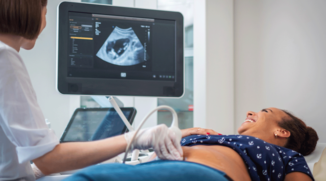
An ultrasound technician—also known as a sonographer—will move a wand back and forth over your belly to evaluate several features of your pregnancy as well as baby’s anatomy. The measurements they take will show if baby’s growth is on track, if you’re having more than one baby and whether or not your due date is accurate, says Erin Clark, MD, chief of division of maternal-fetal medicine at the University of Utah Health. “Knowing that firm due date makes us much more confident to say whether we’re seeing something we’re concerned about or not,” she says.
Typically done in a clinic or hospital setting, your 20-week anatomy scan zeroes in on the health of your pregnancy and baby. Basically, the technician “looks at everything,” says Clark. They’ll measure your little one’s limbs, evaluate the head and spine, check all those budding internal organs and look at baby’s brain anatomy, explains Jennifer Stuck, DO, an ob-gyn with Axia Women’s Health near Philadelphia.
They’ll also look at the amniotic fluid level around baby, and the characteristics of your uterus, ovaries, umbilical cord and placenta, says Clark. If your placenta is located over or near your cervical opening, it could indicate placenta previa, a condition that puts you at a higher risk for vaginal bleeding and warrants additional monitoring, says Stuck.
Identifying baby’s sex
If you haven’t already found out through blood work or a previous ultrasound, the ultrasound technician can usually identify baby’s sex at this point—well, as long as baby cooperates. “We’re at the mercy of the baby. There are many times when the baby is in a position in which everything isn’t as clear as you want it to be,” says Stuck. To dodge any spoiler alerts, tell your technician ahead of time if you don’t yet want to know baby’s sex. (You can also ask them to write it down so you can find out on your own terms.)
Identifying abnormalities
At the 20-week ultrasound, Downs syndrome markers and signs of other anatomical or chromosomal abnormalities, like congenital heart defects, having just one kidney, cleft palate or spina bifida may also be indicated, says Stuck. This can be worrisome, but it also enables you and your medical team to prepare and take steps to improve outcomes.
Heads up: Your anatomy scan won’t be a quick in-and-out appointment. Be ready to hang out on the exam table for as long as it takes for the technician to get the best views of baby—don’t worry, you’re allowed to ask for a bathroom break! The nurses and staff at your practice or hospital will give you specific instructions to prepare you for the 20-week ultrasound in advance. Typically, they’ll ask you to arrive with a full bladder, which works like a window, offering better visualization of baby’s anatomy, says Clark.
You’ll also want to be accompanied by a support person—someone who can provide an extra set of ears for any unexpected news you may get. “It’s good to share in the joy of seeing your baby,” says Stuck. “It’s also good to have that support if they find something you weren’t expecting.”
When making your anatomy scan appointment, be sure to ask when you’ll hear results, says Clark. Will a radiologist or doctor come into the exam room once your scan is finished to talk, or will they read your scan later and call you? Stuck also recommends writing down any updates you’ve given during the appointment, so you can bring questions that pop up directly to your ob-gyn.
Your 20-week ultrasound will last anywhere from 30 minutes to an hour, depending on how stubborn or compliant baby feels that day. (And if you’re expecting multiples, it might take even longer. With twins, an ultrasound at 20 weeks means double the body parts for a technician to evaluate!)
Once you’re reclining on the exam table in the dim room, the technician will dab some gel on your belly and start moving the wand around your stomach and sides, sometimes pushing a bit. It shouldn’t hurt, but it doesn’t quite feel like a massage either.
The tech will be watching images on a screen and typing measurements into a computer. If they’re having trouble seeing something, they may ask you to turn on your side, walk around for a minute or take a bathroom break to pee, says Clark.
In some instances, a transvaginal ultrasound may be necessary. You’ll change into a hospital gown, put your feet into stirrups and have a probe—officially called a transducer—inserted into your vagina. “That allows us to see the low-lying structures a bit easier,” says Clark. “We do what seems reasonable to not make you come back another day for another scan.”
For many moms-to-be, the most trying part of the experience is figuring out what to expect from the ultrasound technician. Some will point out certain things to you on the screen—your baby’s heart, a thumb in their mouth, the length of the legs—while others won’t say much at all. Technicians are certified to know what they’re looking at and how to find everything, but they’re not allowed to diagnose or talk much about what they’re seeing medically, notes Stuck. That’s the job of the doctor who reads the scans later. “Don’t put too much emphasis on [their] facial expressions or reactions during the scan,” says Stuck. Try to relax and enjoy the sweet and reassuring sound of baby’s heartbeat—which, FYI, will likely range between 110 and 160 beats per minute, according to data.
Suffice it to say that the 20-week ultrasound appointment can be exciting, nerve-wracking and exhaustingly long. Your payoff for waiting? A few sweet printed 20-week ultrasound pictures (aka sonograms) of baby that you can keep as mementos—be sure to ask for them before the technician is finished! The image type, either 2D or 3D, will vary based on what technology was used.
The radiologist or doctor employed by the facility you’re at will read your 20-week anatomy scan and analyze the results. They’ll either come into the exam room shortly after your scan, call you with the results later that day or pass them to your primary ob-gyn or for a follow-up call. If the radiology team was able to see everything they needed and everything looks good—and your pregnancy isn’t high-risk—you probably won’t need another ultrasound before delivery, says Stuck.
If the technician wasn’t able to see everything perfectly during your appointment, they may call you to come back for a second scan. “It doesn’t necessarily mean there’s a problem; it means they weren’t able to fully visualize what they needed to see that day,” says Stuck.
If the ultrasound team detects any indication of an abnormality with one of baby’s major organs, your ob-gyn will help you set up a full consultation with a perinatologist (aka a high-risk OB), says Stuck. They’ll come up with a plan for a formal diagnosis that may involve more specialized scans or amniocentesis, and you’ll probably have regular follow-ups throughout the rest of your pregnancy.
Additional screening tests will also be scheduled if your 20-week ultrasound turns up any markers that may indicate a chromosomal issue, such as Down syndrome. “It’s hard to hear that there’s an unexpected finding; it’s easy to get caught up and scared,” says Stuck. “Take a beat, listen to what the physician is saying and know that it may just be a variant of normal.”
The 20-week ultrasound appointment is one of the major landmarks of pregnancy. Try not to stress. Remember, you’ll be able to see your little one on screen—enjoy this sweet moment!
About the experts:
Erin Clark, MD, is the chief of division of maternal-fetal medicine at the University of Utah Health. She earned her medical degree from Mayo Medical School of Medicine.
Jennifer Stuck, DO, is an ob-gyn with Axia Women’s Health near Philadelphia. She received her medical degree from the Philadelphia College of Osteopathic Medicine.
Please note: The Bump and the materials and information it contains are not intended to, and do not constitute, medical or other health advice or diagnosis and should not be used as such. You should always consult with a qualified physician or health professional about your specific circumstances.
Plus, more from The Bump:
Navigate forward to interact with the calendar and select a date. Press the question mark key to get the keyboard shortcuts for changing dates.