


Your Complete Guide to Pregnancy Ultrasounds

The goal of prenatal care is to ensure that both Mom and baby stay safe and healthy as the pregnancy progresses to term. But how exactly do healthcare providers do that? Well, one way is with the use of pregnancy ultrasounds, which help ob-gyns and midwives keep track of baby’s development, Mom’s health and any possible issues that could arise. Here, learn what happens during an ultrasound in pregnancy, how many you might get and when, and why they’re so important for prenatal care.
A pregnancy ultrasound is a prenatal exam—usually done in your doctor’s office—that provides images (called sonograms) of baby and structures of your body, such as your uterus, placenta or ovaries. According to the American College of Obstetrics and Gynecology (ACOG), ultrasounds are conducted using sound waves, which come into contact with the body’s tissues, fluid and bones and then bounce back, providing echoes. These echoes are then received by the equipment used during the exam (more on this below) and turned into a sonogram. These sonograms can offer a 2D, 3D or 4D look at baby in utero.
Types of pregnancy ultrasounds
There are a few different types of ultrasounds in pregnancy to be aware of, according to ACOG:
- Transvaginal ultrasounds are conducted by inserting the transducer into your vagina and moving it side to side to collect sonograms. Early pregnancy ultrasounds are typically conducted transvaginally, since they provide a more accurate view at this stage in baby’s development.
- Transabdominal ultrasound is done when an ob-gyn or ultrasound technician moves a transducer (also called a wand) over your growing bump to get images of baby growing in your uterus.
- 2D ultrasounds are standard ultrasounds (usually conducted transabdominally) that result in two-dimensional images of the developing baby. This is the type of ultrasound most often used in prenatal care.
- 3D ultrasounds are performed as standard transabdominal ultrasounds, but their resulting sonogram offers a three-dimensional look at baby in utero, allowing doctors to get a better look at their development. 3D ultrasounds are typically used following a 2D ultrasound that raises some questions, when a pregnancy is high-risk or when a pregnancy is not moving along as expected.
- 4D ultrasounds—also known as dynamic 3D ultrasounds—are also performed as standard transabdominal ultrasounds, but they offer a three-dimensional look at baby, as well as baby’s movement, similar to a video. While 4D ultrasounds can provide a unique look at baby in utero, they’re usually not medically necessary and more often conducted for “keepsake” purposes.
Fetal ultrasounds offer a safe way for prenatal healthcare providers to keep track of baby’s development and well-being, as well as Mom’s health. Ultrasounds are typically “used throughout pregnancy to determine the number, location, anatomy, position and growth of the baby and the placenta,” says Sherry Ross, MD, an ob-gyn and author of She-ology: The Definitive Guide to Women’s Intimate Health. Period. They can also be used to diagnose any conditions that may arise as the pregnancy progresses. “Ultrasounds can be a helpful tool to identify problems with the baby and pregnancy that can be corrected or minimized if found early on,” Ross says. “It’s one of the best ways to have eyes on a growing baby.” It’s also how providers often confirm a pregnancy to begin with.
How soon can an ultrasound detect pregnancy?
A transvaginal ultrasound can detect a pregnancy as early as five weeks, and a fetal heartbeat around five and a half to six weeks, says Cynthia Flynn, MD, a Florida-based ob-gyn with JustAnswer. According to to Kendra Segura, MD, a California-based ob-gyn, you’ll more likely hear the fetal heartbeat at your 6 week ultrasound, as it’ll be steady by that point and around 110 beats per minute. It’s also important to note that the heartbeat will get stronger over the course of your first trimester as baby’s heart valves and chambers continue to develop.
According to Flynn, in a pregnancy that’s progressing as expected, a typical pregnancy ultrasound schedule involves two scans: one ultrasound in the first trimester and another in the second trimester. However, this number can vary based on the mom-to-be’s individual circumstances. “The frequency of how often an ultrasound is performed depends on when a woman comes to the healthcare provider’s office for their first exam; if she is having any unusual symptoms, including bleeding or pain; or if there are date discrepancies, pregnancy-related problems or anatomical problems for the baby,” Ross says.
An additional ultrasound may also be required if there’s a problem with the initial scan or if it was inconclusive, Flynn notes: “There are so many reasons to do more, but most are due to complications of the pregnancy.”
Pregnancy ultrasounds in the first trimester
According to ACOG, it’s usually too early to see baby’s limbs and organs in detail in the first trimester. Instead, pregnancy ultrasounds at this stage are used to:
- Estimate gestational age (by measuring the length between the crown of baby’s head and their bottom) and due date
- Screen for certain genetic disorders
- Check for twins or triplets
- Check fetal heart rate
- Check for ectopic pregnancy and ensure the pregnancy is viable
Pregnancy ultrasounds in the second trimester
The mid-pregnancy ultrasound occurs between 18 and 22 weeks of pregnancy. According to the American Pregnancy Association, this anatomy scan will check:
- Baby’s development, limbs, anatomy and sex
- Amniotic fluid levels and placenta location
- Signs of abnormalities and markers of Down syndrome, congenital heart disease, neural tube defects, cleft palate and more
- If you’re carrying twins or multiples
Pregnancy ultrasounds in the third trimester
A fetal ultrasound in the third trimester will often look at baby’s growth and positioning, as well as the location of the placenta, the APA says. Flynn adds that an ultrasound in late pregnancy might also be used to monitor any issues, such as fetal abnormalities or fetal growth restriction.
Currently, pregnancy ultrasounds in the third trimester are not a routine part of prenatal care in the United States—and experts disagree on whether they should be. According to one 2020 study, approximately one in 300 women will have a fetal abnormality present during an ultrasound in the third trimester. However, another 2021 review found that data from the past two decades doesn’t make a strong enough case to have a third trimester scan be a routine part of prenatal care.
Ultrasounds in pregnancy have been used for over two decades, and all authorities agree that they’re perfectly safe for both Mom and baby. “Unlike x-rays that use radiation, ultrasounds use sound waves, which do not cause any harm,” Ross says. According to ACOG, there have been no links found between pregnancy ultrasounds and birth defects, childhood cancer or developmental issues later on in life.
However, both ACOG and the FDA note that the long-term potential is still unknown, and pregnancy ultrasound exams should therefore only be performed for medical reasons by qualified healthcare professionals. Both organizations discourage the use of ultrasounds during pregnancy for nonmedical or “keepsake” purposes (such as 3D ultrasounds).
Before your ultrasound, you might be asked to keep your bladder full for a transvaginal ultrasound or empty for a transabdominal ultrasound, Ross says, to help your doctor or technician get clearer imaging. Other than that, there’s not much else you need to prepare, she adds, but it’s best to check in with your doctor’s office for any specific instructions.
So what happens during a pregnancy ultrasound? Transvaginal ultrasounds use a 6-inch probe that’s placed into the vagina to get a look at the uterus, embryo and ovaries. Your doctor or technician will apply a gel lubricant to the wand, have you place your feet into the exam table stirrups and lie back. “It takes around 15 minutes to gather images and take measurements of these structures,” Ross says. You may feel some slight discomfort as the wand is inserted, but “it’s not a painful exam,” she adds.
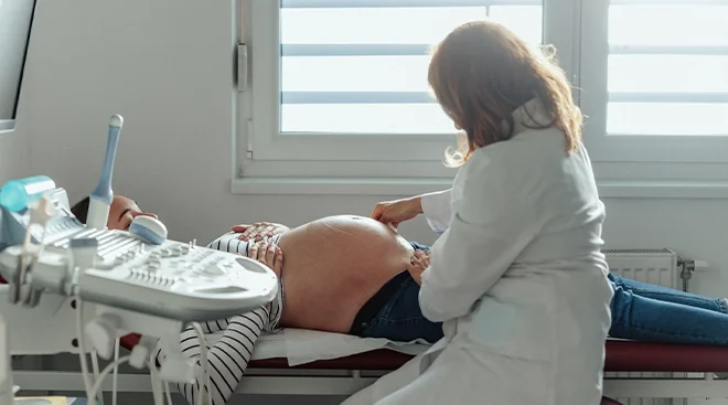
For transabdominal ultrasounds, the probe is much smaller and only used on the exterior of your abdomen to see to the baby, placenta, amniotic fluid and uterus, Ross adds. The technician will apply a lubricant to the wand and glide it over your bump as they capture the necessary images. They may gently press the wand into your abdomen or ask you to shift position so they can get the best angles for clear views, but it shouldn’t be painful. These exams may take 30 minutes to an hour.
If your doctor isn’t the one conducting the ultrasound, don’t be surprised if your ultrasound technician isn’t super forthcoming with details about what they’re seeing on the scans mid-exam. They may explain what they’re taking measurements of or which body part they’re viewing, but their job is to capture the measurements and imaging and pass those to a doctor trained in analyzing sonograms. They then send a report to your prenatal provider, who will walk you through the results. “Expect to need to wait for detailed information until your ob-gyn appointment,” says Flynn.
After the pregnancy ultrasound is over, the technician will wipe the gel from your vagina or abdomen. Both experts agree there generally shouldn’t be any bleeding or needed follow-up care after the exam.
Like so many other aspects of prenatal care, the price tag will vary depending on your insurance, state and where the scan is performed. If your insurance covers prenatal care, it should include at least one scan, Flynn says. Most pregnancy ultrasounds are covered by insurance, Ross adds, but the out-of-pocket costs can range anywhere from $300 to $1,000. It’s also important to note that ultrasounds that are not the standard protocol, like 3D and 4D ultrasounds, may not always be covered by insurance.
Please note: The Bump and the materials and information it contains are not intended to, and do not constitute, medical or other health advice or diagnosis and should not be used as such. You should always consult with a qualified physician or health professional about your specific circumstances.
Plus, more from The Bump:
Cynthia Flynn, MD, is a board-certified ob-gyn based in Florida with over 20 years of experience. She is also an expert with the online platform JustAnswer. She received her degree from the Michigan State University College of Human Medicine.
Sherry Ross, MD, is a women’s sexual health expert and a board-certified ob-gyn at Providence Saint John’s Health Center in Santa Monica, California. She is also the author of She-ology: The Definitive Guide to Women’s Intimate Health. Period. and She-ology, The She-quel: Let’s Continue the Conversation. She earned her medical degree from New York Medical College.
Kendra Segura, MD, MPH, FACOG, is a Los Angeles-based board-certified ob-gyn, as well as an entrepreneur, author, motivational speaker and cast member of Bravo’s hit TV series Married to Medicine: Los Angeles. She earned her medical degree at Ross University School of Medicine in 2011 and completed her residency at Rochester General Hospital in Rochester, New York.
American College of Obstetrics and Gynecology, Ultrasound Exams, October 2021
American Pregnancy Association, Ultrasound Sonogram, 2023
BJOG: An International Journal of Obstetrics & Gynaecology, How often do we identify fetal abnormalities during routine third-trimester ultrasound? A systematic review and meta-analysis, August 2020
Taiwanese Journal of Obstetrics and Gynecology, Third trimester ultrasound. A long-standing debate, May 2021
American College of Obstetrics and Gynecology, Ultrasound Exams, October 2021
US Food and Drug Administration, Ultrasound Imaging, September 2020
Learn how we ensure the accuracy of our content through our editorial and medical review process.
Navigate forward to interact with the calendar and select a date. Press the question mark key to get the keyboard shortcuts for changing dates.