


Sonogram vs. Ultrasound: What’s the Difference?

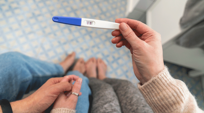
When you’re expecting, getting an ultrasound is one of the most anticipated events of pregnancy. It’s during this exam that you’ll see the first adorable images of your tiny baby—and you’ll probably even get to bring home their very first picture. But if you’ve heard your doctor or someone else refer to the exam as a sonogram, you may be feeling a bit confused about the difference between a sonogram vs. ultrasound. We’re here to clear up the confusion and help you prepare for that exciting doctor’s visit—because we know you’re counting down the days!
So you have a date to “meet” baby on screen? How exciting! One thing you may be wondering as your appointment approaches: Is a sonogram the same as an ultrasound? It’s a legitimate question, and, while the terms are often used interchangeably, they’re actually not synonymous. So what exactly is the difference between sonogram and ultrasound?
The term “ultrasound” refers to the procedure of using sound waves to create an image of an area inside the body, while a sonogram is the actual image that is produced by the ultrasound. Just as a camera captures a photo, an ultrasound exam produces a sonogram image. Basically, the former results in the latter. Still a bit unclear on how it works? Read on for more details about the difference between a sonogram vs. ultrasound—and what you should know before your exam.
An ultrasound is a simple, painless and noninvasive procedure that presents no risk of harm to you or baby. It’s used as a diagnostic exam that allows your doctor to study your growing fetus and track baby’s development at different stages during your pregnancy.
There are two different types of ultrasound examinations typically used during pregnancy:
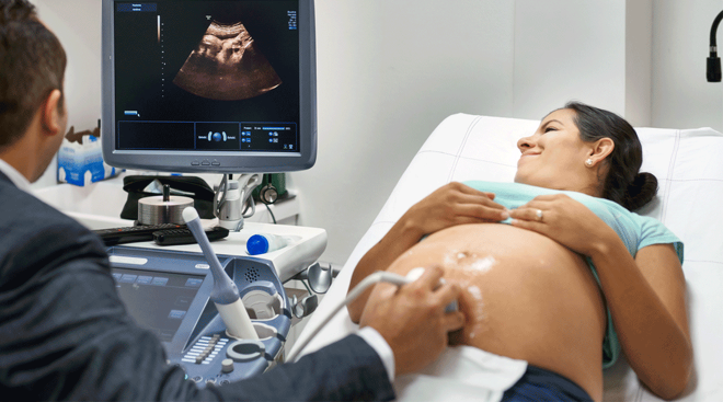
• Transabdominal ultrasound: This is the type of ultrasound you probably envision as you plan for the exciting milestones of pregnancy. During this ultrasound procedure, gel is spread on your belly, and an ultrasound technician, or your doctor, will run a transducer (wand) back and forth over your abdomen to get images of your uterus and baby.
• Transvaginal ultrasound: This exam is often performed in early pregnancy (typically before the eighth week), explains Christian Pope, DO, an ob-gyn at St. Luke’s Hospital in New Bedford, Massachusetts, adding that baby’s heartbeat can usually be detected by transvaginal ultrasound around week six or seven. During the exam, a probe shaped like a wand is lubricated and gently inserted into your vagina. Transvaginal ultrasound is also sometimes used to get a clearer view of the uterus and ovaries.
Your doctor may also use a baby heart monitor or fetal doppler at your regular prenatal appointments beginning around the 12-week mark. This is a handheld device that uses ultrasound technology to check baby’s heartbeat. There’s no visual component.
How does an ultrasound work?
When an ultrasound is performed, internal parts of the body are transformed into images on a screen through the use of a wand or probe. According to Mara Rosner, MD, assistant professor with the department of obstetrics and gynecology at the Johns Hopkins Center for Fetal Therapy, the ultrasound probe transmits high-frequency sound waves. These sound waves bounce off tissue and then back to the transducer probe. The computer then interprets those sound waves to create the image you see on the screen.
What is an ultrasound used for?
With ultrasound technology, your doctor can view your growing fetus and determine things like size, number of fetuses, gestational age and sex. They can also look for any obvious abnormalities, such as spina bifida or cleft palate. Measurements to compare your fetus against others at the same gestational age help ensure that baby is growing and developing properly. By studying baby’s vital organs, like their brain and heart, your doctor can check for proper development and blood flow to those regions.
For the soon-to-be mom, an ultrasound is a great tool that allows a closer look at baby, and it can be an incredibly moving experience to see your little one on screen for the first time. There’s no better bonding experience than realizing you’re growing another human being—and watching them develop with each ultrasound appointment.
If you’re wondering, “Does ultrasound use radiation?,” rest assured that it doesn’t. In fact, because they don’t pose the same risks as x-rays, ultrasounds are becoming increasingly more common in medicine today. Aside from keeping tabs on a pregnancy, doctors can use ultrasound to view things like organs, muscles, heart valves and blood vessels. Ultrasound technology can scan for potential tumors to determine whether or not they’re benign, and it can be used as a guide during procedures like biopsies or amniocentesis (more on that one later).
Ultrasounds during pregnancy
From the moment you see that positive pregnancy test, you’ll likely start wondering, “When do you get your first ultrasound?” It depends on your doctor; many doctors will perform a quick ultrasound at your first pregnancy appointment (which is typically at about 8 weeks). If yours does, don’t expect to see too much this early on—an ultrasound at this stage is mostly for “dating” the pregnancy, or determining a rough due date. Knowing what to expect during each scheduled ultrasound can give you peace of mind. Here’s what to know:
The early pregnancy ultrasound
So how early can an ultrasound detect pregnancy? If you’re at least five to six weeks pregnant, you’ll probably see the beginnings of an amniotic sac and a fetal pole, which will become baby’s body. If your pregnancy is at least six or seven weeks along, you may even get to see a heartbeat.
Of course, there’s always a chance that your pregnancy is a few days earlier than you may think. Because of this high degree of uncertainty, some doctors will skip this early ultrasound altogether—or only recommend it if you’re experiencing bleeding or other troubling symptoms.
These days, there’s little to no preparation for ultrasound necessary. For an early ultrasound, you may be asked to drink water and hold off on urinating for about an hour before your exam. That’s because a full bladder makes it easier to see inside the uterus. Further, Rosner advises that you avoid large meals prior to your exam. “It can make it more uncomfortable to lie flat.”
When you reach what’s called the combined first trimester screening, sometime around weeks 11 to 14, you’ll have the option to have a nuchal translucency screening, or NT scan. Although not quite as exciting as the anatomy scan that you’ll get around 20 weeks, this may be your first “real” glimpse at baby. During this exam, the ultrasound tech will take careful measurements of the back of baby’s neck. “Anything outside of the normal range may be concerning for a genetic problem,” says Pope. With this in mind, your doctor will analyze these measurements, along with blood work, to determine the likelihood of baby having a chromosomal abnormality, such as Down syndrome or trisomy 18. If there’s an increased risk, you can opt for a more invasive test called an amniocentesis. During an amniocentesis, the doctor uses ultrasound to guide a long, thin needle into the amniotic sac. A small amount of fluid is removed and studied to give a more complete picture of baby’s health.
The mid-pregnancy ultrasound
Once you reach the middle of your pregnancy, sometime between weeks 18 and 22, you’ll finally get a chance to have a detailed ultrasound, where you’ll likely be able to see baby from all angles—and get a glimpse at those private parts too! At this ultrasound, determining baby’s sex may be top of mind for you, but it may not be your doctor’s top priority. You’ll likely get an answer to that question (if you want one!), but it’s also key to remember that the technician has an important job to do—so maybe resist the urge to ask for extra pictures. Explains Rosner, “When performing an ultrasound, doctors are looking for anatomical problems and other issues beyond gender. For parents it can be really fun, but it is still a serious medical exam.”
The late pregnancy ultrasound
How many ultrasounds you’ll get during pregnancy depends on your doctor and your situation. Most women have one early on, another at the beginning of the second trimester, and the anatomy scan about halfway through. However, there are a few situations in which additional ultrasounds may be necessary during the third trimester. This late pregnancy exam is often called a biophysical profile, or BPP. “This exam identifies amniotic fluid levels, fetal gross and fine movement and breathing motions,” says Shar La Porte, a certified nurse midwife at Midwifery Care NYC. Here are a few situations that might call for an ultrasound, especially as you approach your due date:
• If you’re having a high-risk pregnancy. In this case, your doctor may want to perform additional ultrasounds to monitor your pregnancy more closely. Advanced maternal age (meaning you’re over age 35, especially if this is your first pregnancy), weakened cervix (which might lead to premature birth), diabetes, high blood pressure and a history of miscarriage are all factors that can cause your doctor to consider your pregnancy high risk.
• If you’re concerned about lack of fetal movements. If you’ve been doing those kick counts diligently but something seems off to you, your doctor will likely perform a late pregnancy ultrasound to ensure that baby is just fine in there. At this stage, an ultrasound may clue your doctor in on potential issues with the umbilical cord.
• If baby is in a breech position. If baby’s not yet in that head-down position, or if you were diagnosed with placenta previa (in which the placenta is covering the uterus), more frequent ultrasounds could be necessary.
• If baby is past 40 to 41 weeks of gestation. The doctor may want to take a peek to monitor fetal well-being.
Whether it’s a scheduled ultrasound or an impromptu check-in, it’s important to know that your technician might not be able to provide answers to your questions. Pope stresses that an ultrasound technician’s job is to take measurements and record ultrasound images, “not to interpret the findings and review with the patient” While this may feel frustrating and disconcerting in the moment, it’s important to stay calm. Oftentimes, this boils down to training and policy. They’ll consult with your doctor who will either come into the exam room to talk to you, or follow-up later.
As previously mentioned, the difference between a sonogram vs. ultrasound is that the ultrasound produces the sonogram. So when your ultrasound exam is over, you’ll probably be handed several pictures of baby growing inside you—the sonogram images. These grainy black-and-white images will become important keepsakes, and you’ll want to show them off to everyone. The word “sonogram” can roughly be translated as “sound writing,” because the images are produced by sound waves.
How to read a sonogram
Here’s where using the words sonogram vs. ultrasound really makes a difference—you can read only the sonogram, not the ultrasound.
If you’re wondering how to read a sonogram, you’re not alone. The images can be difficult to interpret. Ultrasound technology continues to advance, however, and, believe it or not, the images have actually become much clearer over the years. And depending on what your medical facility offers, some ultrasounds allow you to see baby in 3D or even 4D (which means you can see baby moving in 3D) instead of just the flat images produced by 2D ultrasound technology.
Still, if you’re trying to interpret a sonogram image, it’s helpful to be armed with some basic tips. First, the solid colors represent hard tissue and the gray colors, soft tissue. The black areas indicate amniotic fluid. Second, orient yourself by looking for the most obvious parts of baby, like the head or an arm.
You may be wondering how to tell in a sonogram the sex of baby, but you likely won’t have to. Most ultrasound technicians will not only reveal baby’s sex if you ask them to, but will also mark it on the picture and give you a print out. In general, though, the genitalia of baby girls appears as three white lines (which is the clitoris between the two labia). In baby boys, you’ll quite often see a very obvious penis as long as your ultrasound was performed late enough in your pregnancy.
Now that you know the difference between a sonogram vs. ultrasound, you can head into your appointment feeling educated and empowered. Remember, an ultrasound is a safe way to monitor your pregnancy and track baby’s growth. And while seeing baby can be a thrilling experience, it’s, first and foremost, a diagnostic procedure that can bring joyous news or sobering updates. Talk to your doctor if you have questions or concerns. And don’t forget to store baby’s first photo (aka your sonogram printout!) somewhere special.
About the experts:
Shar La Porte, is a certified nurse midwife at Midwifery Care NYC.
Christian Pope, DO, FACG, is an ob-gyn at St. Luke’s Hospital in New Bedford, Massachusetts. He received his medical degree at the Philadelphia College of Osteopathic Medicine.
Mara Rosner, MD, is an assistant professor with the department of obstetrics and gynecology at the Johns Hopkins Center for Fetal Therapy. She received her medical degree at Tulane University School of Medicine in New Orleans, Louisiana.
Please note: The Bump and the materials and information it contains are not intended to, and do not constitute, medical or other health advice or diagnosis and should not be used as such. You should always consult with a qualified physician or health professional about your specific circumstances.
Plus, more from The Bump:
Target Baby Registry
Free $100+ Value Welcome KitFree $100+ Value Welcome Kit
15% Completion Discount15% Completion Discount
Free 1-Year ReturnsFree 1-Year Returns
20+ Exclusive 20% Off
Deals For Mom & Baby20+ Exclusive 20% Off Deals For Mom & Baby1:1 Concierge With
Baby Gear Specialists1:1 Concierge With Baby Gear Specialists

*Subject to availability and Retailer's terms.
We earn commissions from these links.